MRI-Ultrasound Fusion Prostate Biopsy

Medical Review & Verification: Dr. Niti Navanimitkul, M.D.
Board-Certified Urological Surgeon | Advanced Prostate Diagnostics Specialist | M-Trust Urology Clinic
Last Reviewed: July 2026The gold standard diagnostic pathway for prostate cancer precision. An academic comparison of Transrectal (TR) vs. Transperineal (TP) approaches.
What is MRI-Ultrasound Fusion Biopsy?
Traditionally, prostate biopsies were performed as 'blind' systematic procedures (usually 10 to 12 random tissue cores) guided only by standard transrectal ultrasound (TRUS). Because ultrasound cannot visualize most prostate tumors, this blind method missed up to 30-40% of significant cancers, leading to false negatives [1].
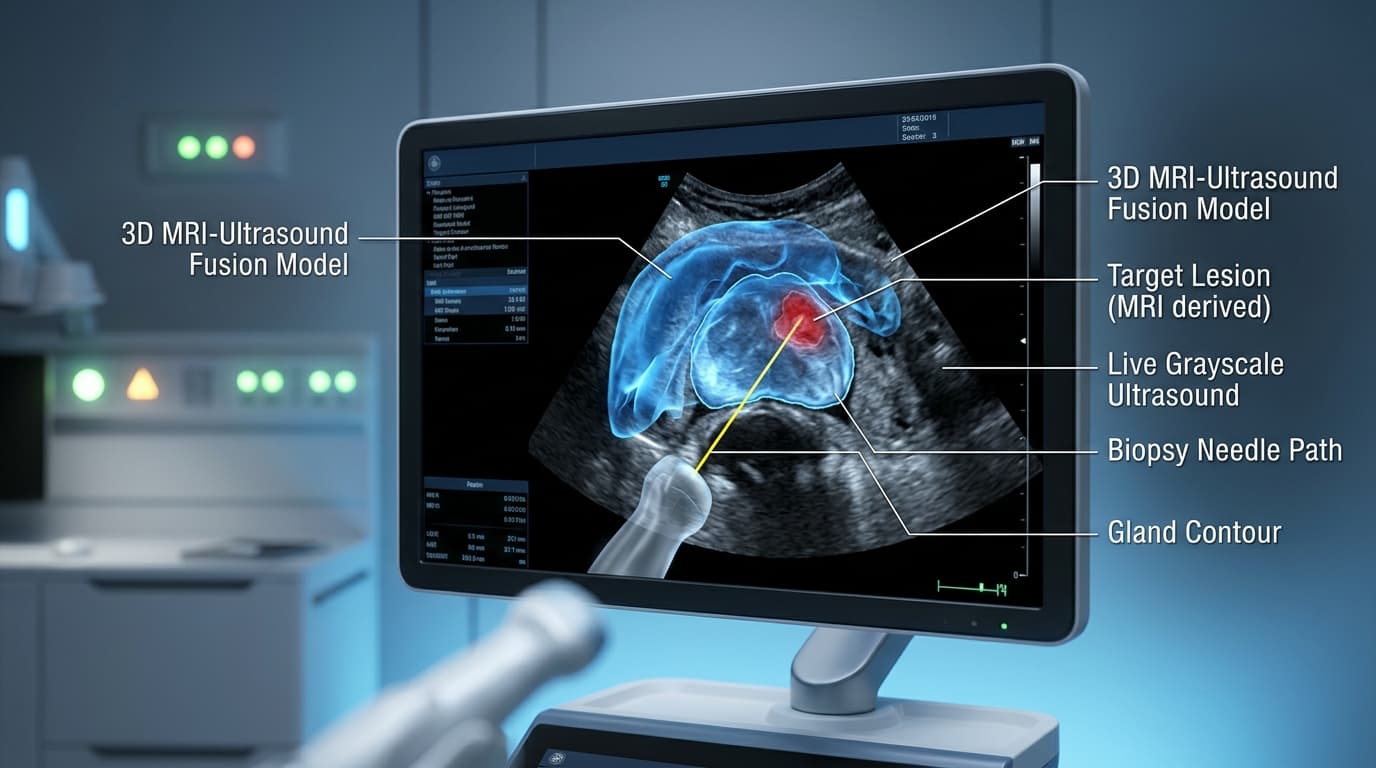
The **MRI-Ultrasound Fusion Biopsy** solves this by importing a patient's pre-procedure multiparametric MRI (mpMRI) scan into advanced mapping software. The software reconstructs a 3D model of the prostate and overlays it directly onto live ultrasound images during the biopsy. This allows the urologist to guide the biopsy needle to precise suspicious target lesions (PIRADS 3-5) with millimeter accuracy [2].
TR vs. TP Biopsy Approaches
There are two primary approaches to access the prostate gland for biopsy. The pathway chosen significantly affects patient safety, infection risk, and accuracy [3]:
Approach Breakdown & Clinical Analysis
1. Transrectal (TR) Approach
The biopsy needle is inserted through the rectal wall directly into the prostate gland. - **Infection Risk:** Passing the needle through the bacteria-laden rectum into the prostate carries a higher risk of urinary tract infection or life-threatening bloodstream infection (sepsis) of 1-2%, despite prophylactic antibiotics. - **Access Limitations:** The entry angle makes it difficult to biopsy the apical (bottom) and anterior (front) zones of the prostate, potentially missing significant cancers located there [4].
2. Transperineal (TP) Approach
The biopsy needle is inserted through the sterile perineal skin (the area between the scrotum and anus). - **Near-Zero Sepsis Risk:** Because the skin is disinfected beforehand and the rectum is bypassed entirely, the sepsis rate drops below 0.1%. - **Unrestricted Targeting:** The needle travels parallel to the prostate, allowing complete access to all areas, including the anterior and apical zones [5].
How the Procedure is Performed
The biopsy is performed as an outpatient procedure under local perineal block, sedation, or general anesthesia [6]:
1. Image Registration
Pre-biopsy mpMRI images are fused with real-time ultrasound images to map target areas.
2. Targeted Biopsy
The urologist takes 2-4 targeted cores from the suspicious PI-RADS 3-5 lesions visible on the 3D map.
3. Systematic Biopsy
An additional 10-12 standard cores are taken from other regions to check for microscopic disease.
Pre-Procedure Patient Preparation
Following these steps reduces bleeding and ensures a smooth procedure [7]:
1. Medication Adjustment
Stop all blood thinners (aspirin, clopidogrel, anticoagulants) 5-7 days before the biopsy.
2. Antibiotic Prophylaxis
Take prescribed oral antibiotics or receive intravenous antibiotics prior to the procedure as directed.
3. Bowel Prep & Fasting
A fleet enema is recommended on the morning of the procedure. Fast for 6 hours if sedation or general anesthesia is used.
Side-by-Side Comparison: Transrectal (TR) vs. Transperineal (TP) Biopsy
An academic comparison of the clinical safety and efficacy profiles [8]:
Efficacy and Limitations of MRI Fusion Biopsy
⊕ ข้อดี (Pros)
- ✓Provides the highest diagnostic accuracy, reducing cancer missed rates by over 30%.
- ✓Saves patients from unnecessary repeat biopsies due to targeted precision.
- ✓Accurately differentiates between low-grade and clinically significant prostate cancer.
- ✓Transperineal approach virtually eliminates the life-threatening risk of sepsis.
- ✓Delivers a precise target map crucial for focal therapies or radiation planning.
⊖ ข้อจำกัด (Cons)
- ✗Requires high clinical expertise and specialized fusion imaging software.
- ✗Requires a high-quality pre-procedure mpMRI scan.
- ✗Costs are higher than standard blind/random ultrasound biopsies.
Who is a Candidate for MRI Fusion Biopsy?
Recommended diagnostic pathways according to international guidelines [9]:
- ▹Patients with elevated or rising PSA levels: Or abnormal findings on a Digital Rectal Exam (DRE).
- ▹Patients with a PI-RADS 3, 4, or 5 lesion: Identified on a recent prostate mpMRI.
- ▹Patients with previous negative biopsies: But who have persistently high or rising PSA levels.
- ▹Patients planning focal therapies (e.g. HIFU, NanoKnife): Where exact tumor boundaries must be mapped.
Clinical Efficacy & Cancer Detection Rates
Key performance metrics supported by global clinical studies [10]:
Higher Detection Rate
Increased detection of clinically significant cancers compared to blind biopsy.
Sepsis Rate (TP)
Almost zero risk of severe blood infections with transperineal access.
Targeting Accuracy
Highly reliable needle placement inside the MRI-detected target lesion.
Post-Procedure Timeline
What to expect during the healing phase [11]:
- •Pathology Report: Results are processed by a pathologist and available in 5-7 business days.
- •Hematuria: Mild blood in your urine (pinkish color) or minor rectal spotting is normal for 3-5 days.
- •Hemospermia: Blood in semen (brownish discoloration) can persist for 4-6 weeks and is harmless.
Post-Biopsy Care & Recovery Guidelines
Instructions to prevent complications and speed up recovery [12]:
- •Complete Antibiotics: Finish your entire course of prescribed antibiotics without interruption.
- •Stay Well Hydrated: Drink 2.5 to 3 liters of water daily to flush the bladder and minimize dysuria.
- •Avoid Pelvic Friction: Refrain from heavy lifting, strenuous exercise, running, or cycling for 7 days.
- •Watch for Warnings: Go to the emergency room immediately if you develop a high fever, chills, inability to urinate, or heavy bleeding.
Related Topics
Academic References & Medical Literature
- Grummet JP, et al. 'Transition to Transperineal Biopsy: Sepsis Control and Quality Outcomes.' BJU International, 2020; 126(3):361-368. doi:10.1111/bju.15082.
- Xiang J, et al. 'Transrectal vs. Transperineal MRI-Ultrasound Fusion Guided Prostate Biopsy: A Systematic Review.' Journal of Urology, 2021; 206(3):584-593. doi:10.1097/JU.0000000000001859.
- Mottet N, et al. 'EAU-EANM-ESTRO-ESUR-SIOG Guidelines on Prostate Cancer.' European Association of Urology Guidelines, 2024.