Focal Therapy for Localized Prostate Cancer

Medical Review & Verification Details: Dr. Niti Navanimitkul, M.D.
Board-Certified Urologist | Specialist in Minimally Invasive Urological Surgery & Advanced Prostate Treatments
Last Reviewed: July 2026An innovative, tissue-sparing treatment that destroys only the cancer tumor, preserving potency and urinary continence with high precision under Multiparametric MRI (mpMRI) guidance.
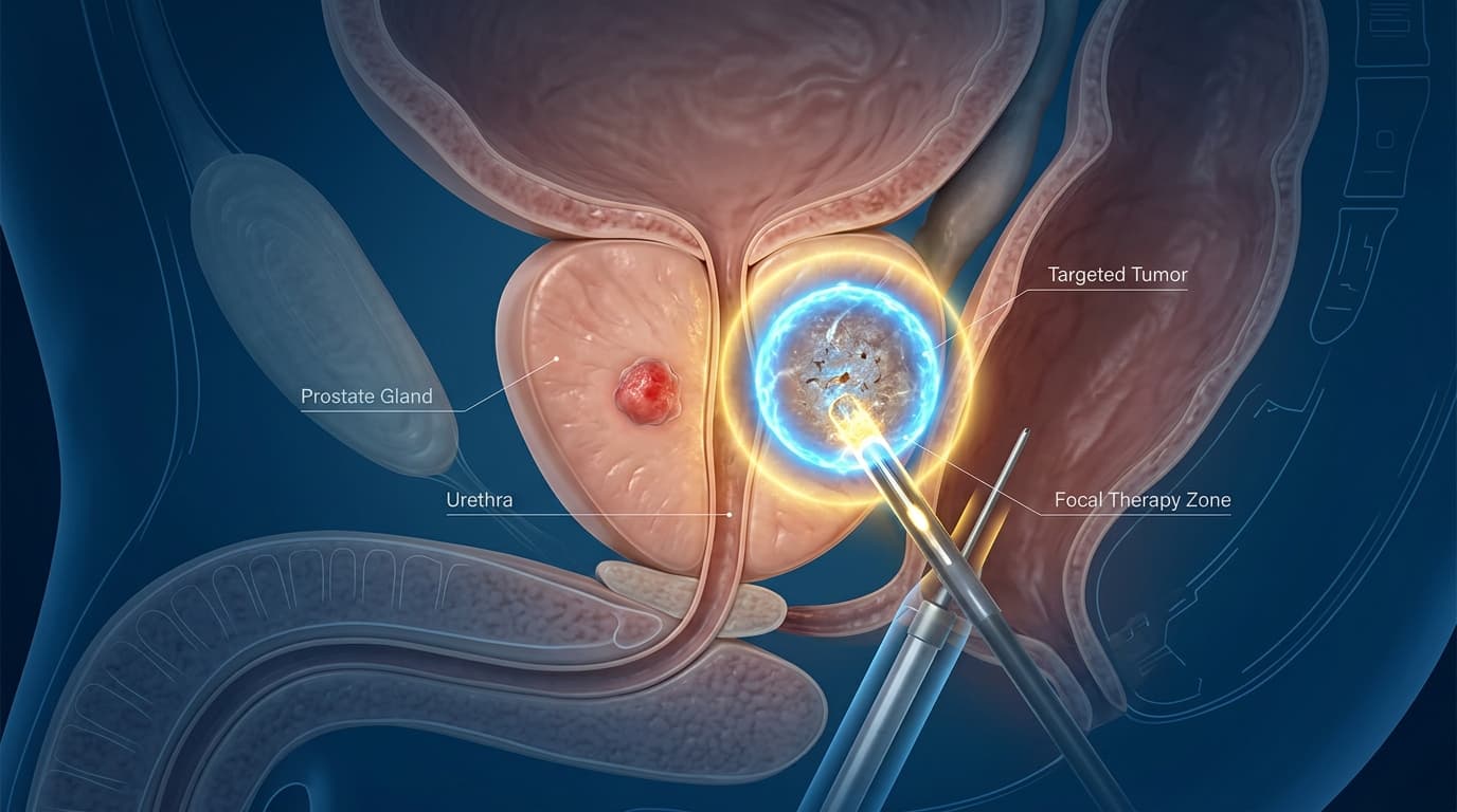
What is Focal Therapy?
Focal Therapy is an emerging treatment paradigm for patients diagnosed with localized, low-to-intermediate risk prostate cancer. Rather than treating or surgically removing the entire prostate gland—as with Radical Prostatectomy or Whole-Gland Radiation—Focal Therapy specifically targets and destroys only the distinct areas of cancer within the gland [1].
Advancements in Multiparametric Magnetic Resonance Imaging (mpMRI) enable highly accurate mapping of the tumor location. Under MRI guidance or MRI-ultrasound fusion, urologists can deliver targeted energies to destroy cancer cells while sparing healthy prostate tissue, the neurovascular bundles (controlling erections), and the urinary sphincter (preventing incontinence) [2].
The Index Lesion Concept
Prostate cancer is often multifocal, meaning multiple small tumors can exist in the prostate. However, clinical and pathological research shows that the 'index lesion'—the largest tumor with the highest Gleason score—almost exclusively drives the progression of the disease and metastatic spread. Successfully ablating this index lesion along with a safe treatment margin controls the cancer as effectively as radical whole-gland therapies, while sparing the patient from major treatment morbidity [3].
Ablation Technologies in Focal Therapy
Focal Therapy uses various energy sources to ablate cancer cells based on the size, location, and shape of the tumor [4]:
1. HIFU (High-Intensity Focused Ultrasound)
Delivers focused ultrasound waves transrectally, converging at a precise focal point to generate temperatures of 80-90°C. This induces coagulative necrosis of the cancer cells without injuring the intervening rectal wall or surrounding structures. Approved by the FDA and referenced in AUA guidelines [5].
2. Cryotherapy (Cryoablation)
Uses thin needle-like probes inserted percutaneously through the perineum into the tumor under ultrasound guidance. Circulation of argon gas drops temperatures to below -40°C, creating an 'ice-ball' that ruptures cell membranes via crystallization and induces vascular thrombosis in the tumor [6].
3. Irreversible Electroporation (IRE / NanoKnife)
A non-thermal ablation method that delivers short, high-voltage electrical pulses (up to 3,000V) between probes placed around the tumor. This creates permanent, nanoscale pores in the cell membranes (electroporation), triggering natural apoptosis (cell death) while preserving the surrounding extracellular matrix, vascular scaffolds, and nerve fibers [7].
Pre-operative Diagnostic Workup
To ensure that focal therapy is safe and effective for you, the clinical evaluation must follow strict protocols [8]:
Multiparametric MRI (mpMRI)
A high-resolution MRI scan scored by PI-RADS (scores 3 to 5) to precisely locate, map, and measure the boundaries of the index lesion.
MRI-Ultrasound Fusion Biopsy
Fusing real-time ultrasound with the pre-op MRI to direct biopsy needles precisely into the target lesion, supplemented by systematic biopsy of the remaining prostate lobes.
Risk Stratification
Ensuring low-to-intermediate risk disease: PSA levels below 15 ng/mL, clinical stage ≤T2a, and Gleason Score of 6 (3+3) or 7 (3+4 or 4+3).
Metastatic Workup (PSMA-PET/CT)
If clinically indicated, a highly specific PSMA-PET scan is performed to verify that the cancer is localized strictly to the gland and hasn't metastasized [9].
Comparison of Early-Stage Prostate Cancer Treatments
An analytical medical comparison between current treatment choices for localized prostate cancer [10]:
Who is a Candidate for Focal Therapy?
Careful patient selection is paramount to achieving oncological outcomes comparable to radical treatments. Candidates typically meet the following criteria:
- ▹Localized Prostate Cancer: Clinically staged as T1c to T2a (cancer confined to the gland).
- ▹Low-to-Intermediate Risk: Gleason Score of 6 or 7 (Grade Group 1 or 2).
- ▹PSA Level Below 15 ng/mL: With a distinct tumor lesion visible on mpMRI.
- ▹Unilateral Disease: Tumor limited to one side of the prostate or a clearly defined dominant index lesion.
- ▹Quality of Life Focused: Patients highly motivated to preserve erectile function and urinary continence.
Clinical Outcomes & Success Statistics
Aggregated statistical data from major peer-reviewed clinical trials demonstrating the safety and efficacy of focal ablation [12]:
Continence Preservation
Almost all patients recover immediate, complete bladder control without leaking.
Erectile Function Preservation
Maintains sexual health, erectile firmness, and quality of life compared to surgery.
5-Year Local Cancer Control
High success rates in local control for well-selected patients.
Post-procedure Surveillance Protocol
Because the remaining prostate tissue is left intact, patients must follow a structured monitoring calendar to guarantee long-term cancer control and catch any recurrence early [11]:
- •Month 1: Post-procedure evaluation, assessment of urinary voiding patterns and initial potency baseline.
- •Months 3, 6, 9, 12 (Year 1): Serum PSA tests every 3 months to monitor PSA kinetics and define the PSA Nadir (lowest point). A rapid rise in PSA warns of potential residual disease.
- •Months 6 - 12: Repeat multiparametric MRI (mpMRI) to evaluate the treated ablation zone and ensure the remaining gland is clear.
- •Month 12: A confirmatory fusion biopsy of the treated zone and any newly suspicious MRI findings to confirm complete oncological clearance.
- •Year 2+: PSA checks every 6 months, and follow-up mpMRI scans every 1-2 years as directed by your urologist.
Recovery & Post-procedure Guidelines
Focal therapy is a minimally invasive outpatient procedure. Most patients experience a rapid recovery by adhering to the following guidelines [13]:
- •Temporary Catheterization: Temporary prostate tissue swelling occurs. A urinary catheter is left in place for 3 to 7 days to prevent urinary retention.
- •Mild Discomfort: You may experience mild burning during urination or pelvic pressure for the first 1-2 weeks. This is managed with oral analgesics and antispasmodics.
- •Pink-tinged Urine: Traces of blood in your urine or semen are normal during the first few weeks and will resolve on their own.
- •Restricted Activities: Avoid strenuous exercises, bicycling, or heavy lifting for 2-3 weeks to prevent delayed prostate bleeding.
Related Topics
Academic Medical References & Peer-Reviewed Journals
- Valerio M, et al. 'New and Established Technology in Focal Therapy for Prostate Cancer: A Systematic Review.' European Urology, 2016; 69(1):119-128. doi:10.1016/j.eururo.2015.08.026.
- Coleman JA, et al. 'Focal Therapy for Prostate Cancer: AUA/SUO Guideline.' Journal of Urology, 2023; 209(6):1096-1104. doi:10.1097/JU.0000000000003437.
- Nyein A, et al. 'The Index Lesion Concept in Localized Prostate Cancer.' Pathology and Oncology Research, 2020; 26(3):1429-1435. doi:10.1007/s12253-019-00712-4.
- Stabile A, et al. 'Medium-term Outcomes of Focal Therapy for Prostate Cancer: A Systematic Review.' European Urology Focus, 2022; 8(4):918-927. doi:10.1016/j.euf.2021.08.004.
- National Institute for Health and Care Excellence (NICE). 'High-intensity focused ultrasound for prostate cancer.' Interventional procedures guidance IPG673, London: NICE, 2020.
- Vargas HA, et al. 'The Role of Multiparametric Magnetic Resonance Imaging in Focal Therapy Planning and Response Assessment for Prostate Cancer.' Oncology, 2018; 32(8):390-396.
- Schatloff O, et al. 'Irreversible Electroporation (NanoKnife) for Localized Prostate Cancer: 3-Year Follow-up.' BJU International, 2021; 128(S3):25-32. doi:10.1111/bju.15582.